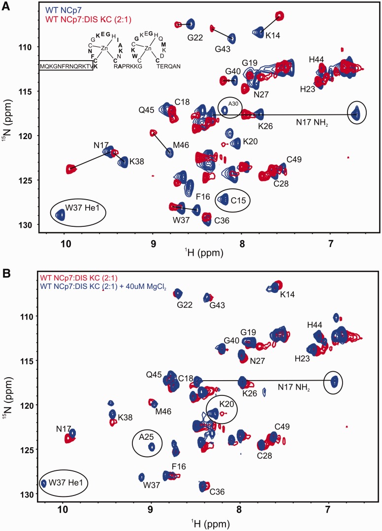
Figure 5.
Structural characterization of wild-type NCp7 binding to the DIS kissing complex. (A) An overlay of the 15N-1H HSQC spectrum of the wild-type NCp7 (blue) and its complex with the DIS kissing complex (red). The backbone amide resonances are labeled as well as the side chain indole NH group (He1) of W37 and the side chain amino resonances (NH2) of N17 are shown with a solid line connecting them. Resonances that are broadened beyond detection on binding the RNA are circled, and shifted resonances are indicated with solid lines. The secondary structure of NCp7 is drawn with residues showing significant chemical shift perturbation in bold. The N-terminal residues for which amide resonances are not observed in the HSQC spectrum are boxed. (B) An overlay of the 15N-1H HSQC spectrum of the wild-type NCp7 bound to the DIS kissing complex (red) and the same complex after addition of 40 µM of MgCl2 (blue). The residues whose amide proton resonances either reappear or sharpen on addition of 40 µM of MgCl2 are labeled.

