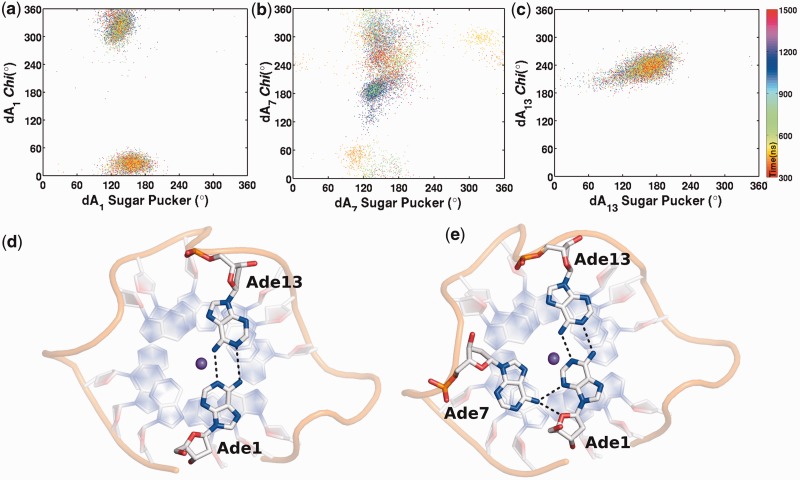
Figure 4.
Time development of A:A base pair and A:A:A triad on the terminal quadruplex. Scattergram of glycosidic chi angle versus sugar pucker angle of (a) Ade1, (b) Ade7 and (c) Ade13. (d) The syn conformation of Ade1 and anti conformation of Ade13 facilitates reverse Watson and Crick A:A base pairing on terminal quartet. (e) Ade7 flips to syn conformation and interacts with Ade1 to form A1:A7:A13 triad. The K+ ion is represented as purple sphere.

