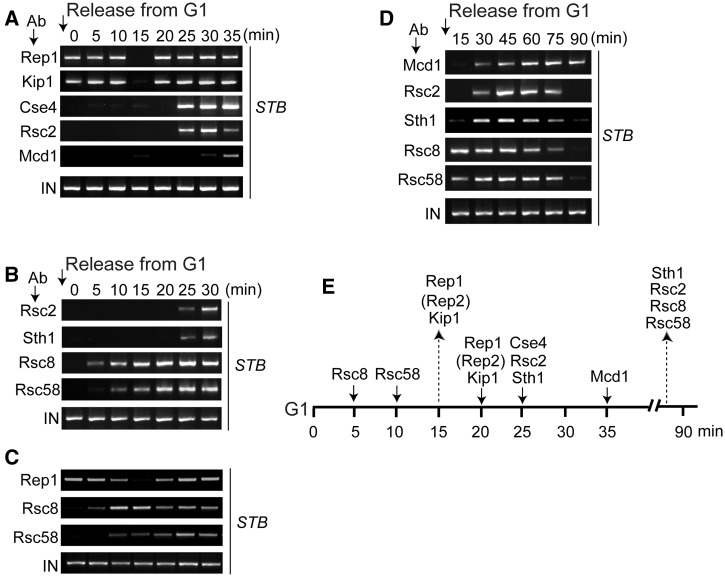
Figure 3.
Temporal sequence of events at STB during the assembly and disassembly of the plasmid partitioning complex. ChIP assays were performed in cells arrested in G1 (time zero), and at every 5 min time point after their release into the cell cycle. In each panel, the top row indicates the reference protein that was paired with each of the others in individual assays. (A) Rep1 was not epitope-tagged; Mcd1 and Cse4 harbored HA3- and Myc12-epitopes, respectively, and the others Myc13-epitope. (B) Rsc2 harbored Myc13-epitope; the others HA3-epitope. (C) Rep1 was not epitope-tagged; Rsc8 and Rsc58 were tagged with Myc-13 epitope. (D) Mcd1 harbored HA3-epitope; the others Myc13-epitope. In each individual assay, after splitting samples into equal portions, the reference protein was immunoprecipitated in one set and the test protein in the other. Rep1 was immunoprecipitated using an antibody to the native protein. Each of the other proteins was immunoprecipitated using an antibody to the epitope tag harbored by it. (E) The cumulative results from A–D are placed along the time line for plasmid segregation. Rep2, placed in parentheses, was not analysed in the present assays. Based on previous analyses, it is expected to follow the Rep1 pattern. The strains for these assays were MJY3162 to MJY5042 (Supplementary Table S1).

