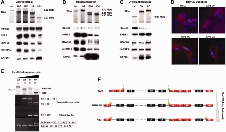
Figure 6.
Titin size is determined by the relative ratio of Rbm20 to other splicing factors. (A) Upper panel shows titin proteins separated on an sodium dodecyl sulphate–agarose gel. The left lane is from an Hm adult LV, and the rest are Wt from fetal (F) and postnatal heart aged in days (d). Lower panel shows western blots. Sample loads were balanced for Rbm20 content. Results show that during development, the size reduction in titin protein is accompanied by increased ratios of Rbm20 to other splicing factors. (B) Similar developmental changes in the ratios of Rbm20 to other splicing factors occur in the skeletal muscle tibialis anterior (TA). The Rbm20 to splicing factor ratios increase with age, whereas the titin size decreases. (C) Tissue specific Rbm20 and titin isoform expression in LV, TA and longissimus dorsi (LD). Sample loads were balanced for the splicing-related factors. The results demonstrate that the ratio of Rbm20 to other splice factors is inversely related to titin isoform size. (D) Characterization of Rbm20 speckles during development and between muscle tissues. Two speckles per nucleus were observed in all the indicated tissues. d, day(s). (E) Titin isoforms and splicing pathways from different muscle tissues. Titin isoforms from different tissues are shown in the upper panel. TA 2 and N2BA N1 correspond to the cooperative repression between exons 50 and 70. TA2 is smaller than N2BA N1 because only the latter contains protein coded by exon 49, which accounts for an additional 0.1 MDa of protein mass (21). N2B results from the alternative 3′ ss pathway (shown by exons 50–219). The 5′ ss of exon 70 is always active for splicing, but the 3′ ss of exon 70 can be repressed in some titin mRNA in Ht LV and Wt LV (bottom panel). The unspliced 3′ ss of exon 70 cannot be found in Wt LD and Wt TA. (F) A schematic diagram demonstrating the Rbm20/splicing factors ratio–dependent extension of Rbm20-repressed region between exons 50 and 70 and the consequence of each situation. As the Rbm20/splicing factors ratio increases from top to bottom, the Rbm20-repressed region extends from exons 51–70 to exons 50–70. When Rbm20/splicing factors ratio is high enough like in Ht and Wt LV, we found the splicing of the 5′ ss of exon 50 to downstream alternative 3′ ss, for example the 3′ ss of exon 219. The 5′ ss of exon 50 can splice with many other alternative 3′ ss (refer to Figure 5B); they are not shown in this diagram. The dotted lines denote the continuous exons (and introns if repressed by Rbm20) that are not shown.

