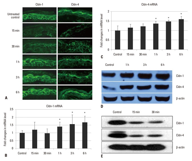
Fig. 1.
Effect of acute permeability barrier disruption on the expression and localization of TJ proteins, Cldn-1 and Cldn-4 in murine epidermis. Skin samples were taken at 15 min, 30 min, 1 h, 3 h, and 6 h after tape-stripping. Frozen sections (5 µm) were immunostained with Cldn-1 and Cldn-4 primary antibodies (Zymed Laboratories, San Francisco, CA, USA) and an FITC conjugated donkey anti-rabbit IgG, secondary antibody (Santa Cruz, CA, USA) and examined by confocal microscopy. Magnification ×400 (A). The levels of mRNA for Cldn-1 (B) and Cldn-4 (C) were determined using real-time PCR and normalized to that of β-actin. Cldn-1 and Cldn-4 protein expression was determined by western blot analysis of tape-stripped murine epidermis (D and E). β-actin was used as a loading control. *p<0.05 for Cldn-1 and Cldn-4 mRNA of tape-stripped epidermis compared with that of untreated control. TJ, tight junction; Cldn, claudin. FITC, fluorescein isothiocyanate; PCR, polymerase chain reaction.

