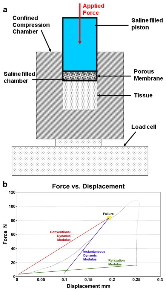
Fig. 1.
Apparatus and method used to quantify pseudo-dynamic modulus in myometrial and leiomyoma surgically obtained tissue samples. a: Schematic of the experimental confined compression apparatus with a porous membrane (40 micron pore size). A 5% constant displacement uniaxial load was applied to the myometrial and leiomyoma tissue. The confined compression chamber was smooth, rigid, and impermeable. b: Representative Force versus Displacement graph for a single leiomyoma specimen.

