
Fig. 3.
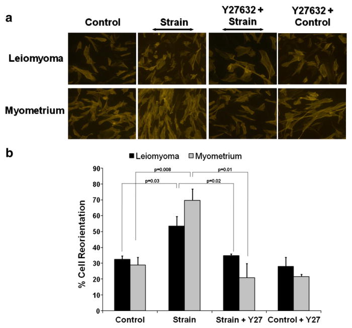
Response of myometrial and leiomyoma cells to cyclic uniaxial strain. a: Cytoimmunofluorescent images of leiomyoma and myometrial cells exposed to either no strain (control) or to 8.9% uniaxial cyclic strain (Strain) for 18 h at 1 Hz. Cells were cultured with or without pre-treatment of the ROCK inhibitor, Y-27632 (Y-27) (10 μM) for 30 min prior to strain or no strain (control). Actin stress fibers and nuclei were visualized by staining for Alexa Fluor-546 Phalloidin and DAPI, respectively. b: Quantitative computerized morphometric measurements of cellular reorientation in response to uniaxial strain with, or without, pre-treatment of Y-27632 (Y-27) for leiomyoma (black bars) or myometrial cells (grey bars). Results are shown as the percentage of cells aligned at 90°+/−30° relative to the direction of the applied strain. Data represent a mean of three independent experiments with a minimum of 45 cells measured per condition. Angular differences between unstrained and strained leiomyoma and myometrial cells differed significantly (p<0.05).