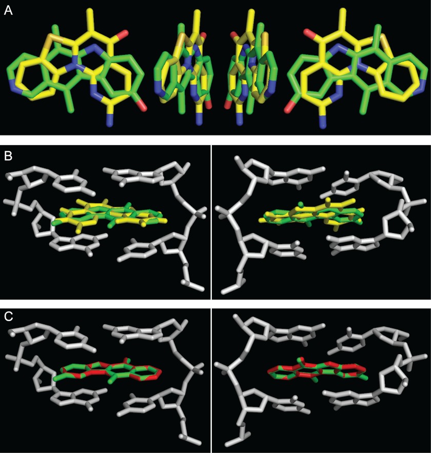
FIGURE 12.
Structural alignment of 9HE, GQC-Qi, and CX-5461 hydrophobic cores and their docking position in the DNA strand. A, shown is a three-dimensional superimposition of aromatic cores of 9HE (carbon atoms are in green) and CX-5461 (carbon atoms are in yellow). B, shown is a three-dimensional superimposition of 9HE (green) and CX-5461 (yellow) in the complex with d(GpC) (gray). Only aromatic cores are shown. Shown are the views from the major (left) and minor (right) grooves. C, shown is a three-dimensional superimposition of 9HE (green) and GQC-Qi (red) in complex with d(GpC) (gray). Only aromatic cores are shown. Shown are the views from the major (left) and minor (right) grooves.

