FIGURE 6.

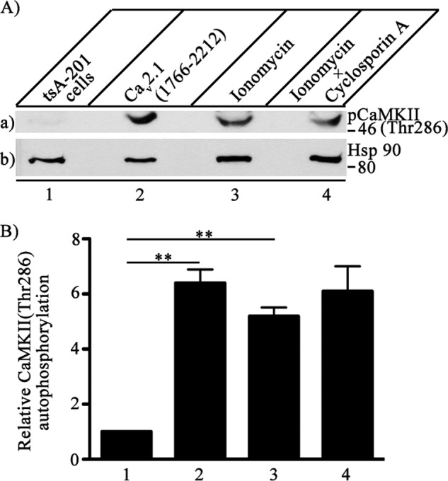
Activation of autophosphorylation of CaMKII by binding to CaV2.1 channels. CaV2.1(1766–2122) was expressed in tsA-201 cells, and autophosphorylation of endogenous CaMKII was measured with anti- phospho-CaMKII(Thr-286). A, left to right, untreated control tsA-201 (lane 1), cells transfected with C-terminal domain of Cav2.1 (1766–2212) (lane 2), cells treated with 5 μm ionomycin to allow Ca2+ entry for 15 min before lysis (lane 3), and cells treated with 5 μm ionomycin and additionally with cyclosporin A to inhibit the protein phosphatase calcineurin (lane 4). a, autophosphorylation of CaMKII was assayed by immunoblotting with anti- phospho-CaMKII(Thr-286). b, the same blot after stripping and re-probing with anti-Hsp90 to show equal loading of cell lysates. B, quantitation of relative CaMKII autophosphorylation using anti-phospho-CaMKII(Thr-286) under the indicated conditions (means ± S.E.; **, p < 0.01 by Student's t test; n = 3).
