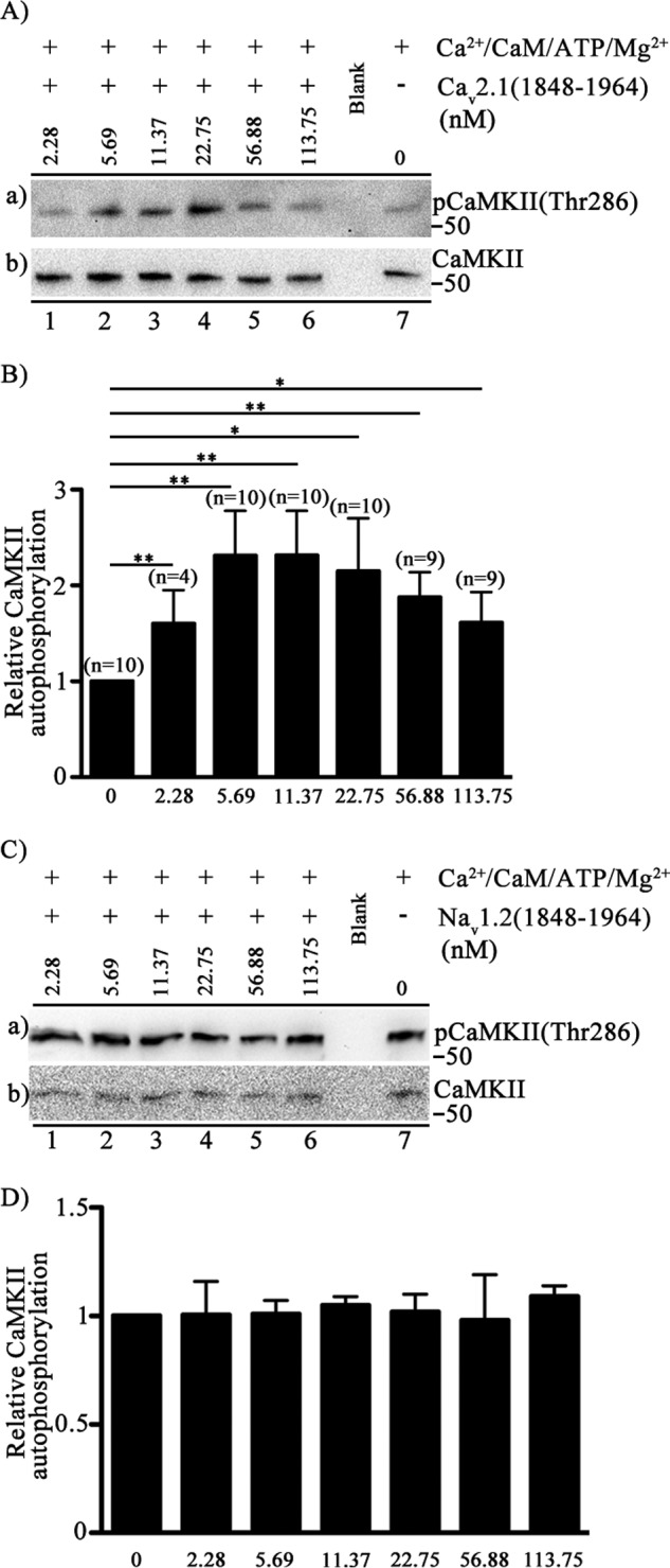
FIGURE 7.

Activation of autophosphorylation of CaMKII by binding of CaV2.1(1848–1964). A, autophosphorylation of CaMKII was carried out by incubation in the presence of Ca2+ (0.6 μm), CaM (2.2 μm), ATP (32 μm), Mg2+(2.3 mm), CaMKII (20 nm) and the indicated concentrations (0–113.75 nm) of Cav2.1 (1848–1964). Free Ca2+ was precisely controlled. Left to right (lanes 1–6), the concentration of CaV2.1(1848–1964) channel peptide ranging from 2.28 nm to 113.75 nm. Extreme right (lane 7), the control in the absence of CaV2.1(1848–1964). a, the blot was probed with anti-phospho-CaMKII(Thr-286). b, the same blot after stripping and re-probing using anti-CaMKII. B, quantitation of relative CaMKII autophosphorylation using anti-phospho-CaMKII(Thr-286) under the indicated conditions (mean ± S.E.; *, p < 0.05; **, p < 0.01 by Student's t test). C, CaMKII autophosphorylation in the presence of NaV1.2(1848–1964)-GST is shown. Autophosphorylation of CaMKII was carried out by incubation in the presence of Ca2+ (0.6 μm), CaM (2.2 μm), ATP (32 μm), Mg2+(2.3 mm), CaMKII (20 nm), and the indicated concentrations (0–113.75 nm) of NaV1.2(1848–1964). Free Ca2+ was precisely controlled. Left to right (lanes 1–6), concentration of NaV1.2(1848–1964) channel peptide ranging from 0 to 113.75 nm. Extreme right (lane 7), control in the absence of NaV1.2(1848–1964). a, the blot was probed with anti- phospho-CaMKII(Thr-286). b, the same blot after stripping and re-probing using anti-CaMKII. D, the quantification of relative CaMKII autophosphorylation using anti-phospho- CaMKII(Thr-286) under the indicated conditions (mean ± S.E.; not significant by Student's t test; n = 3).
