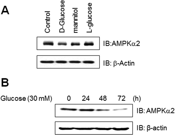
FIGURE 1.
AMPKα2 down-regulated in C2C12 cells cultured under high glucose conditions. A, C2C12 cells were stimulated for the indicated agents, such as d-glucose (30 mm), l-glucose (30 mm), and mannitol (30 mm) for 6 days. The cell lysates (20 μg) were analyzed via Western blotting for anti-AMPKα2 antibody. The anti-β-actin antibody was a protein loading control. B, C2C12 cells were stimulated for the indicated times with 30 mm high glucose. The cell lysates (20 μg) were analyzed via Western blotting for anti-AMPKα2 antibody. The anti-β-actin antibody was a protein loading control.

