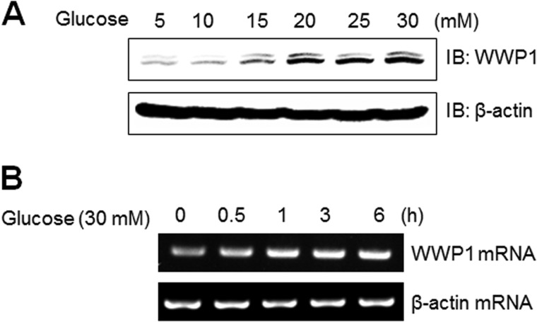
FIGURE 5.
WWP1 increased in high glucose conditions. A, C2C12 cells were stimulated for the indicated doses of glucose for 48 h. The cell lysates (20 μg) were analyzed via Western blotting for anti-WWP1 antibody. The anti-β-actin antibody was a protein loading control. B, total mRNA was prepared for these cells after 30 mm glucose incubation, and RT-PCR was conducted using specific WWP1 primers. The PCR product was then gel-run in 1% agarose, and visualized in UV. β-Actin was employed as a positive control.

