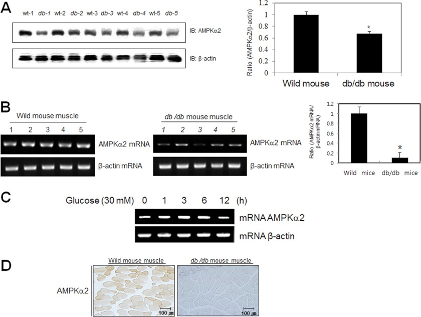
FIGURE 6.
AMPKα2 down-regulated in db/db mouse. A, Western blot analysis of quadriceps muscles of wild type and db/db mice. The tissue lysates (40 μg) were analyzed via Western blotting for anti-AMPKα2 antibody. The anti-β-actin antibody was a protein loading control. Densitometry analysis (n = 5) of values are means ± S.E. values of the ratios of densities (AMPKα2/β-actin). *, p < 0.05 versus wild type mouse. B, RT-PCR analysis of quadriceps muscles of wild type and db/db mice. Total mRNA was prepared for these mice, and RT-PCR was conducted using specific AMPKα2 primers. The PCR product was then gel-run in 2% agarose, and visualized in UV. β-Actin RNA was employed as a positive control. Densitometry analysis (n = 5) of values are means ± S.E. values of the ratios of densities (AMPKα2 mRNA/β-actin mRNA). *, p < 0.05 versus wild type mouse. C, total mRNA was prepared for these cells after 30 mm glucose incubation, and RT-PCR was conducted using specific AMPKα2 primers. The PCR product was run in 1% agarose gel, and visualized in UV. β-Actin was employed as a positive control. D, histochemistry of the quadriceps muscle of wild type and db/db mice. Each cryosection was stained with anti-AMPKα2 antibody.

