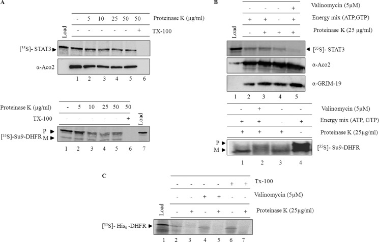
FIGURE 2.
In vitro import of STAT3 into isolated mitochondria. A, in vitro import of STAT3 and Su9-DHFR was performed for 60 min as described under “Experimental Procedures.” After import, samples were treated with increasing concentrations of proteinase K (lanes 2–6) either in the absence (2–5) or presence of Triton X-100 (lane 6). After inhibition of proteinase K with PMSF (1 mm), samples were re-isolated and resolved on SDS-PAGE (P, precursor; M, mature). B, import of STAT3 (top panel) and Su9-DHFR (bottom panel) was examined in the absence of membrane potential by incubating mitochondria with valinomycin (5 μm) for 5 min (top panel, lane 5; bottom panel, lane 2) or by excluding the external energy source in the form ATP, GTP, and succinate (top panel, lane 4; bottom panel, lane 3) in the import reaction. C, import of His6-DHFR was carried out as described under “Experimental Procedures,” and the samples were treated with proteinase K (25 μg/ml) and analyzed by phosphorimaging.

