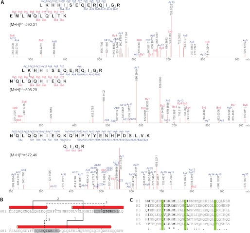
FIGURE 6.
Identification of Plugin cross-linking sites. A, CID-MS/MS analyses and corresponding fragment ion series for three specific peaks identified by mass spectrometric analysis of cross-linked Plugin-C tryptic digest. B, location of cross-linking sites within Plugin-C sequence. C, alignment of α-helices in Plugin C-terminal domain. The positions of known cross-linked Gln residues are highlighted in green, conserved or highly similar residues are shown in bold.

