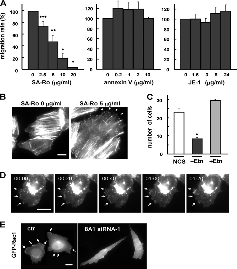
FIGURE 7.
Inward translocation of cell surface PE is involved in cell migration. A, migratory activity of CDC50A-overexpressing cells was examined by the transwell assay, in the presence of PE-binding probe (SA-Ro), the PS-binding probe (annexin V), or anti-PC monoclonal antibody (JE-1) at the indicated concentrations. The number of migrating cells without addition of probes was normalized to a 100% migration rate. Values are means ± S.D. from three independent experiments. B, CDC50A-overexpressing cell line 13 was washed and incubated in serum-free Ham's F-12 containing 0.1% BSA and 700 μg/ml hygromycin B in the presence (left) or absence (right) of 5 μg/ml SA-Ro for 3 h at 37 °C. Cells were then fixed and stained for the distribution of F-actin using TRITC-labeled phalloidin. Arrowheads indicate the spike-like membrane protrusions. The scale bar indicates 10 μm. C, R-41 cells were cultured in Ham's F-12 medium containing 10% newborn calf serum (NCS). For ethanolamine depletion, R-41 cells were cultured in medium containing 10% dialyzed newborn calf serum (−Etn) for 48 h. For restoration of PE synthesis, R-41 cells were cultured in medium containing 10% dialyzed newborn calf serum and 20 μm ethanolamine-HCl (+Etn) for 48 h. The transwell assay was performed as described above. Values are means ± S.D. from three independent experiments. *, p < 0.05; **, p < 0.01. D, localization of ATP8A1 in migrating cells. The cell line stably expressing GFP-ATP8A1 was serum-starved for 24 h and then incubated in the presence of 10% newborn calf serum. After serum stimulation for 50 min, images from GFP-positive cells were recorded every 20 s for 1.5 min. Representative cells are shown at 0, 20, 40, 60, and 80 s. Arrows indicate the highly motile ruffling membranes. E, CHO cell line stably expressing GFP-Rac1 was transiently transfected with control siRNA (ctr) and siRNA-1 (8A1 siRNA-1) and was observed after 3 days. Arrows indicate the distribution of Rac1 at the membrane ruffles.

