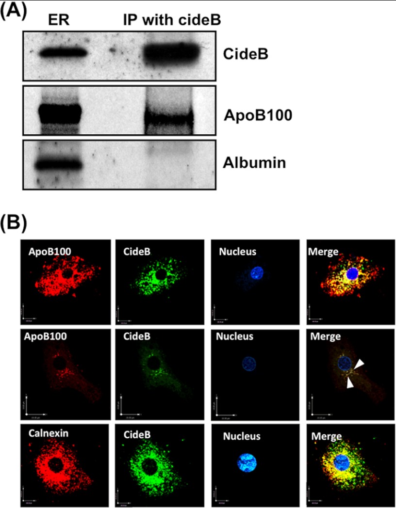
FIGURE 3.
CideB interacts with apoB100 but not with albumin. A, ER (250 μg of protein) were solubilized in 2% (v/v) Triton X-100 and incubated with anti-goat CideB antibody (10 μg) for 4 h at 4 °C. Anti-goat IgGs bound to agarose beads were added and incubated overnight at 4 °C. Immunocomplexes bound to agarose beads were isolated and washed 10 times with ice-cold PBS to remove unbound proteins. Protein sample was separated by SDS-PAGE (8–16% gel) and probed with anti-CideB, anti-apoB100, and anti-albumin antibodies. IP, immunoprecipitation. B, CideB co-localizes with apoB100 in hepatic ER. Primary hepatocytes were double-labeled with either CideB (FITC, green) and apoB100 (Texas Red, red) (upper and middle panels) or CideB (FITC, green) and calnexin, an ER marker (Texas Red, red) (lower panel). In the middle panel, we used higher magnification and less saturation of both channels. Arrowheads in the middle panel show punctate vesicular staining in the VTVs. The nucleus is stained with DAPI (blue). Merged figures show co-localization of CideB with apoB100 and calnexin.

