
FIGURE 12.
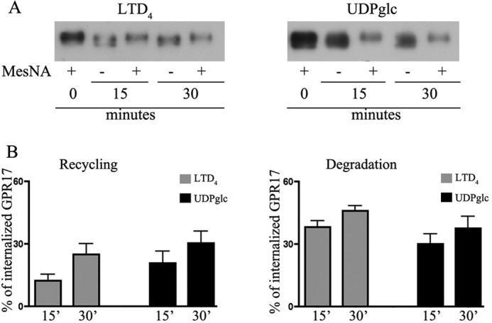
Recycling and degradation of GPR17 after agonist-induced endocytosis. Oli-neu cells after cell surface labeling with sulfo-NHS-SS-biotin at 4 °C were incubated at 37 °C with UDP-glucose (100 μm, UDPglc) or LTD4 (50 nm). After 15 min, cells were cooled at 4 °C, incubated with MesNA to remove biotin from the remaining cell surface receptors, and solubilized (time 0) or further incubated in normal medium. At selected times, cells were either solubilized (−) or re-incubated with MesNA to remove biotinylated receptors re-exposed to the cell surface. In A aliquots (200 μg) of cell extracts were incubated with streptavidin beads, and the proteins bound to streptavidin were analyzed by Western blotting with antibodies against GPR17. In B, graphs represent the quantitative analysis of GPR17 recycling and degradation, which is reported as a percentage of the amount of receptor internalized after 15 min at 37 °C with LTD4 or UDP-glucose; the values are the mean of four independent experiments ±S.E.