
FIGURE 4.
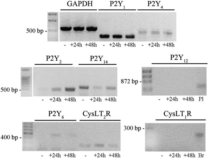
Analysis of purinergic and CysLT receptors in Oli-neu cells. Reverse transcription-PCR amplification was performed using specific primers (see “Experimental Procedures”) and 1 μg of RNA extracted from Oli-neu cells cultured in Sato medium alone (−) or in medium supplemented with CD (+) for 24 or 48 h. RNA from mouse brain (Br) and mouse platelets (Pl) was used as positive controls as indicated under “Experimental Procedures.” GAPDH was used as an internal standard. The final products were separated on an agarose gel (5 μl of the reaction mixtures were loaded on each lane). The bands revealed by ethidium bromide staining corresponded to the predicted sizes of the amplified DNA fragments.