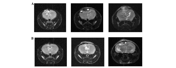
Figure 4.
Serial magnetic resonance imaging (MRI). (A) A tumor was noted in the cortex of the right hemisphere 4 weeks after intracranial inoculation of 1×10⁶ NCI-H358 cells (left). The tumor size progressively increased and compressed the brain parenchyma at 8 weeks (middle) and 12 weeks (right) after the inoculation of control mice. (B) In mice treated with rapamycin before inoculation, the tumor formed and thrived relatively slowly. Left, 4 weeks; middle, 8 weeks; right, 12 weeks.

