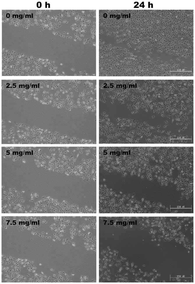
Figure 4.
Effect of MTE on HUVEC migration. After treatment with the indicated concentrations of MTE for 24 h, the migration pattern of the HUVECs was observed using phase-contrast microscopy. The images were obtained at a magnification of ×100. Images are representative of three independent experiments. MTE, Marsdenia tenacissima extract; HUVEC, human umbilical vein endothelial cell.

