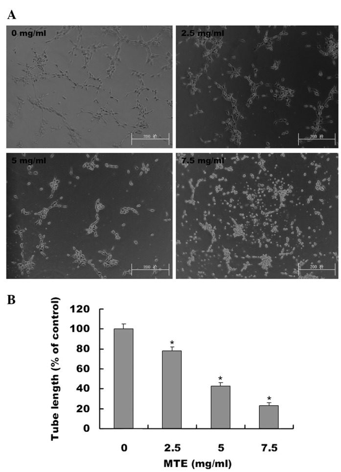
Figure 5.

The effect of MTE on tube formation in HUVECs. (A) HUVECs were harvested and suspended in medium containing various concentrations of MTE. The harvested cells were then seeded in basement protein (ECMatrix, Millipore)-coated plates and incubated for 9 h at 37°C. The development of network-like, tube structures was observed by phase-contrast microscopy. The images were obtained at a magnification of ×100. Images are representative of three independent experiments. (B) The total length of the capillary-like tubes was measured and normalized to the control. Data shown are the mean ± SD (error bars) from three independent experiments. *P<0.01, vs. control cells. MTE, Marsdenia tenacissima extract; HUVEC, human umbilical vein endothelial cell.
