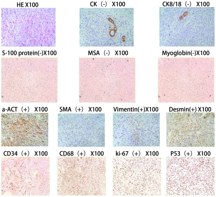
Figure 2.
Light microscopy. The tumor cells exhibited embryonic mesenchymal differentiation and lacked epithelial characteristics. Moreover, the tumor cells demonstrated fusiform and star shapes, which have undetermined boundaries and clear heteromorphism. Changes in the interstitial mucus and the periodic acid-Schiff (PAS)-positive eosinophilic body, both inside and outside the tumor cells, were evident. The remnant hepatic cells and hyperplastic bile duct are visible in the boundaries of the tumor, and there were no changes in the hepatic cirrhosis. The immunohistochemistry results are as follows: Broad spectrum creatine kinase (CK)(−), cytokeratin 8/18(CK8/18)(−), S-100(−), MSA(−), myoglobin(−), SMA foci(+), a-ACT foci(+), desmin foci(+), vimentin foci(+), CD34(+), CD68(+), Ki-67(+) and P53(+).

