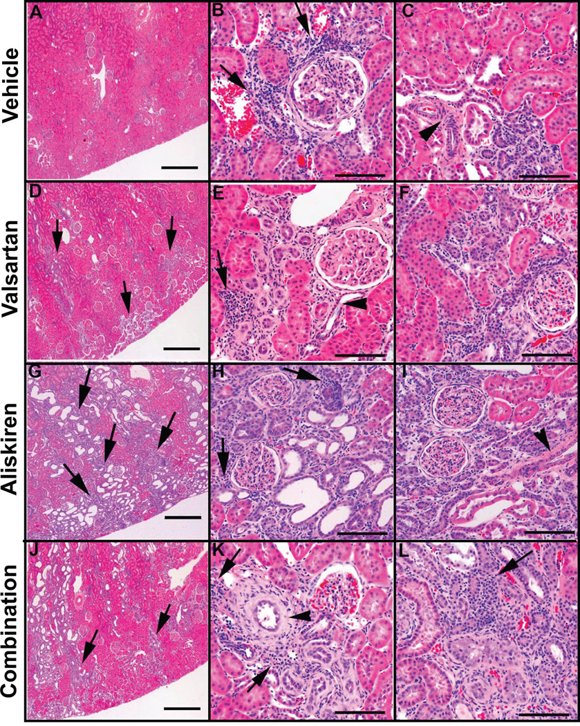
Figure 3.
Hematoxylin-eosin stained sections of renal cortical tissue obtained from representative vehicle (panels A, B, and C), valsartan (panels D, E, F), aliskiren (panels G, H, and I), and valsartan/aliskiren (panels J, K, and L)-treated mRen2.Lewis hypertensive rats. At low magnification vehicle treated mRen2.Lewis hypertensive rats shows an overall conserved renal architecture (A), whereas valsartan (D and G) -treated mRen2.Lewis rats show abnormal areas (arrows) with tubular proliferation, periglomerular and peritubular fibrosis and tubular dilatation, more extensive in the aliskiren or the combined treatment groups (J). Lymphocytic infiltrates surrounding tubules and vessels are observed in all groups (arrows in panels B, E, H and K). Examples of periglomerular fibrosis and signs of glomerulosclerosis are shown in panels B, E, F, H and I. Areas consisting of high nuclear to cytoplasm ratio, peritubular and periglomerular fibrosis and disorganized tubular proliferation (basophilic areas) are small in the vehicle group (B and C) but are more frequent and extended in the valsartan (E and F), aliskiren (H and I) and combined treatment group (K and L). Most of the arteries/arterioles are fairly conserved (C, E and I, arrowheads) but some are very abnormal denoting a hypertrophic response with thickening of the media (panel K, arrowhead). Scale bar: A, D, G and J: 500 µm; B, C, E, F, H, I, K and L: 100 µm.

