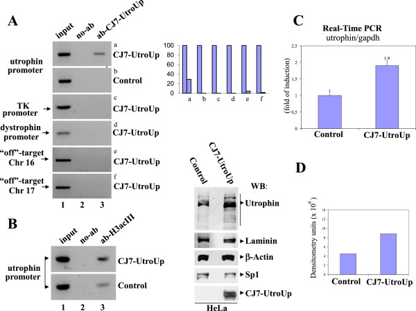
Figure 4.
The binding and upregulation of the endogenous utrophin gene. A: (Left) The ChIP assay was performed in transfected HeLa cells (with either an empty control vector or with the CJ7-UtroUp expression vector) using myc-tag monoclonal antibody/protein G-agarose beads. The immunoprecipitates from each sample were analysed by PCR performed using primers specific for the human utrophin promoter “A”. PCRs of the promoters of the unrelated thymidine kinase and dystrophin genes and PCRs of specific regions on the chromosomes 16 and 17 were included as controls. A sample representing the linear amplification of the total input chromatin (input) was included (lane 1). (Right) Histogram shows quantification of ChIP-PCR bands presented as fraction of the input. B: ChIP was performed in transfected HeLa cells (with either the CJ7-UtroUp expression vector or with an empty control vector) using anti-acetylated histone H3 rabbit polyclonal antibody/protein G-agarose beads. Immunoprecipitates from each sample were analyzed by PCR performed using primers specific for human utrophin promoter “A”. C: The analysis by real-time PCR of the utrophin gene mRNA expression in HeLa cells transfected with the CJ7-UtroUp or control vectors. The ratio between the utrophin and GAPDH gene expression is shown as means ± S.D. from three independent experiments that were performed in triplicate. D: (Left) Western blot analysis of total protein lysates from HeLa cells transfected with either empty vector (Control) or with the CJ7-UtroUp expression vector. The membrane was incubated with the indicated antibodies. (Right) Histogram shows utrophin expression evaluated by densitometric analysis.

