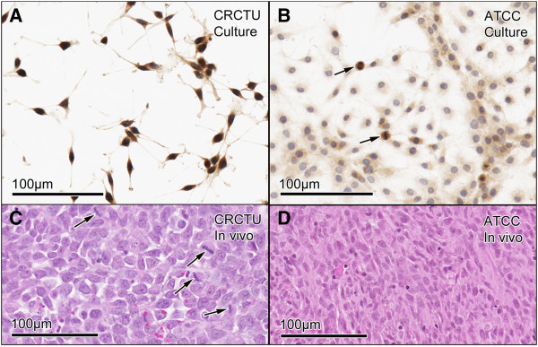
Figure 1.

Tumour cell morphology in vitro and in vivo. (A) CRCTU Walker 256 cells in culture stained for cytokeratin 18, showing spicular appearance with deeply-stained nuclei. (B) ATCC Walker 256 cell in culture stained for cytokeratin 18 showing flattened cells with a large cytoplasmic component and lightly stained nuclei and mitotic figures (arrows). (C) CRCTU Walker 256 tumour cell in vivo, 9 days following internal carotid artery injection, stained with haematoxylin and eosin showing large nuclei and scanty cytoplasm with many mitotic figures (arrows). (D) ATCC Walker 256 tumour cell in vivo, 10 weeks following internal carotid artery injection, showing smaller nuclei and abundant elongated cytoplasmic component, giving the section an eosinophilic appearance.
