Figure 6.
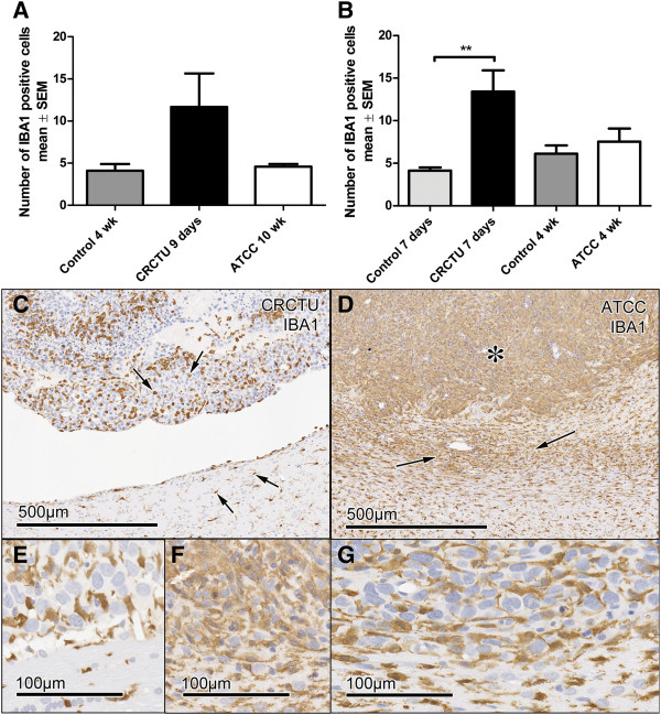
IBA1 immunoreactivity. (A) The average number of IBA1 positive cells /0.0678 mm2 of the cortex in animals injected with culture medium, CRCTU or ATCC Walker 256 breast carcinoma cells into the internal carotid artery. (B) The average number of IBA1 positive cells /0.0678 mm2 of the striatum following direct inoculation of culture medium, CRCTU or ATCC Walker 256 cells into the brain; **p<0.01. (C) IBA1 immunostained brain 9 days following internal carotid artery inoculation with CRCTU Walker 256 cells, arrows showing labelled cells dispersed between cancer cells and in the peritumoral area. (D) IBA1 immunostained brain section showing extensive labelling within the tumour mass (asterisk) and in the peritumoral area (arrows) 10 weeks following ATCC Walker 256 breast carcinoma cell injection into the internal carotid artery. (E) Higher magnification IBA1 immunostained section 9 days following CRCTU Walker 256 internal carotid artery injection. (F) Higher magnification IBA1 immunostained section 10 weeks following ATCC Walker 256 internal carotid artery injection. (G) IBA1 immunostained section 7 days following direct inoculation of CRCTU Walker 256 inoculation showing increased immunoreactivity in the peritumoral area along with IBA1 labelled cells within the tumour mass.

