Figure 3.
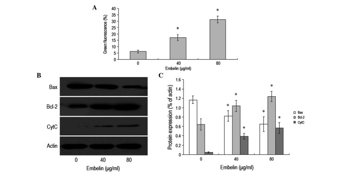
Embelin induced MCF-7 breast cancer cell apoptosis via the mitochondrial pathway, following treatment of MCF-7 cells with different concentrations of embelin (0, 40 and 80 μg/ml) for 48 h. (A) JC-1 dye flow cytometry was used to analyze the change in the mitochondrial membrane potential. (B) Western blot analysis was used to determine the expression levels of Bax, Bcl-2 and cytochrome C proteins inside the cytoplasm. (C) The western blot analysis results were further analyzed using Gel-Pro Analyzer 4.0 software. *P<0.05 vs. control. Results are representative of three independent experiments.

