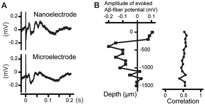
Figure 3. Electrically evoked intracortical field potentials recorded in the rat primary somatosensory cortex (acute measurements).
(A) Simultaneous recordings using a nanowire-based electrode and a microwire electrode glued together and implanted 400 µm below the cortex surface (averaged over 32 sweeps). (B) Depth profile of evoked Aβ-fiber potential (filled boxes) recorded by the nanowire-based electrode (plotted for each depth as the peak-valley amplitude, with an onset latency between 10 ms and 20 ms after the stimulation) and correlation coefficients (filled circles) calculated for measurements performed simultaneously with the nanowire-based electrode and the microwire (calculated for the measured data sets of time interval up to 0.43 ms after the stimulation). The measurements show that the neuronal signal is primarily recorded with the nanowire-based sensing part and that the nanowire-based electrode provides acute in vivo recordings that are comparable to conventional microelectrodes.

