
Figure 5. SEM images of the nanowires modified sensing site.
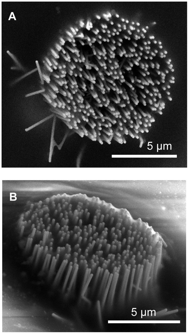
The site image presented after a single implantation (A) and after multiple implantations (B) into rat cortex. The same nanowires-based electrode before any implantation can be seen in Figure 1B. Some tissue deposition on the probe after multiple implantations can be seen in Figure 5B.