
Figure 6. RPV-Sa infection blocks IFNα-induced phosphorylation of Jak1/Tyk2.
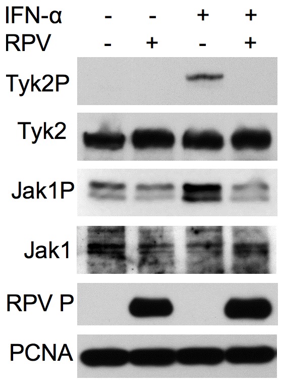
A549 cells were infected with RPV-Sa at a MOI of 5 or left uninfected. Eighteen hours post-infection cells were mock treated or treated with 1000 IU/ml of IFNα for 15 minutes, lysed in SDS-PAGE sample buffer containing protease and phosphatase inhibitors and the levels of phosphorylated Jak1 and Tyk2 were analyzed in Western blots using rabbit anti-Jak1P and rabbit anti-Tyk2P antibodies respectively. The blots were also probed with rabbit anti-Jak1, rabbit anti-Tyk2 and rabbit anti-RPV P protein (MB18) to analyze the total Jak1 and Tyk2 and to confirm RPV infection. The primary antibodies were detected with peroxidase-labelled anti-mouse or anti-rabbit IgG antibody. PCNA levels served as loading control.