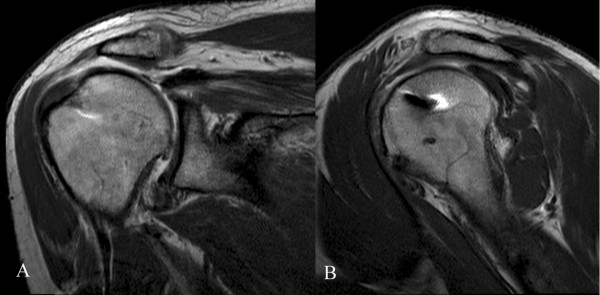
Figure 4.

Coronal (A) and oblique sagittal (B) MRA views of the operated shoulder of a 48-year-old man (SR group), at 38-month follow-up, showed a type II tear of the rotator cuff, with a thin supraspinatus tendon (4.5 mm thick), leakage of the contrast medium, and superior migration of the humeral head. Despite the radiological findings, clinical results were good with 74 points at CMS and 12 points at SST.
