
Figure 2.
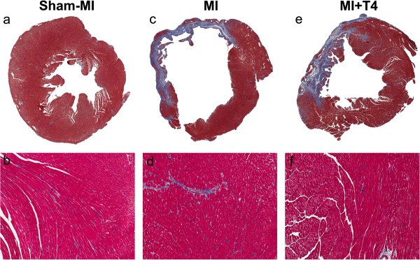
Changes in collagen deposition in the LV non-infarcted area at 8 weeks post-MI. Transverse myocardial sections, Masson’s Trichrome staining. (a, c, d) Left ventricle and septum, 0.5x magnification. (b, d, f) Enlarged image from septum, 10x magnification.