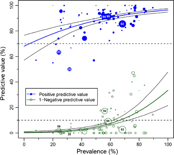
Figure 1.

Predictive values of coronary computed tomography angiography. The population-averaged predictive values (based on published studies and their summary results) are shown as blue (positive) and green (negative) curves with their 95% confidence intervals based on the published studies (circles). The negative predictive values are shown as 1-negative predictive values to avoid overlap with the positive predictive value curve. The number of patients included in the published studies is indicated by the size of the circles. The dashed lines indicate the predefined minimal predictive values on the y-axis (90% for negative and 70% for the positive predictive values). Such analyses are limited because only the published predictive values in the studies and the calculated prevalences (but not the pretest likelihoods [7-9]) are available. Thus, individual patient data (IPD) from a collaborative meta-analysis can overcome these limitations and draw more meaningful conclusions about the clinical utility of diagnostic tests. Image reproduced from: Schlattmann P, Schuetz GM, Dewey M: Influence of coronary artery disease prevalence on predictive values of coronary CT angiography: a meta-regression analysis. Eur Radiol 2011, 21(9):1904–1913 [12].
