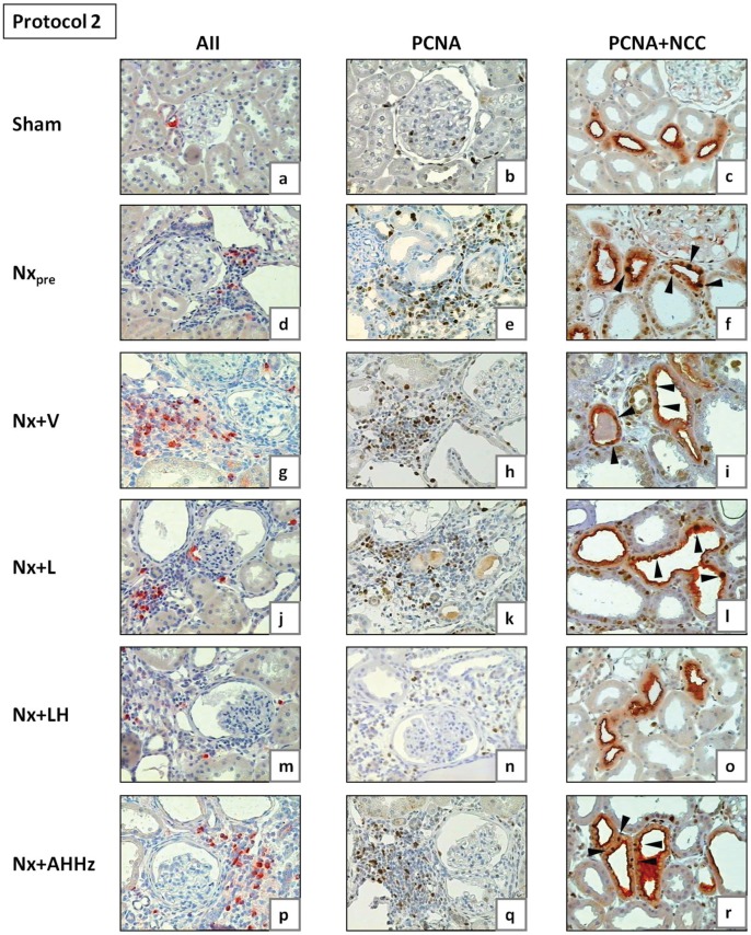
Figure 11. Representative microphotographs of renal tissue obtained for Nxpre (120 days after renal ablation) and for all other groups (210 days after renal ablation) in Protocol 2.
AII, tubulointerstitial cells staining positively for AII; PCNA, proliferating-cell nuclear antigen; NCC, sodium-chloride cotransporter, specific for distal convoluted tubule (DCT). Arrowheads in Figs. 11c, f, i, l, o and r (double staining for PCNA and NCC) indicate examples of PCNA-positive cells in DCT.

