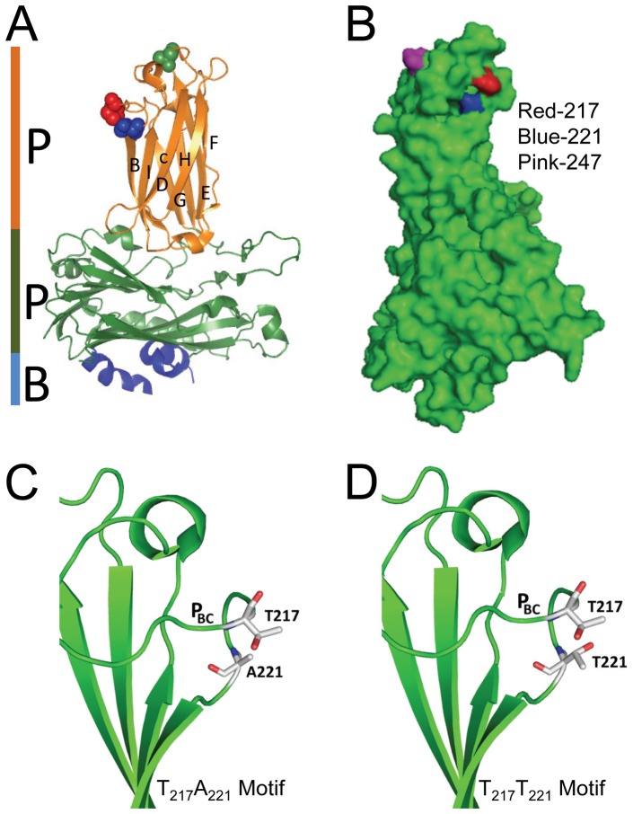
Figure 11. Crystal structure of the VP2 subviral particle (SVP).
A) Shows the P, S and B domains of the VP2-SVP while spheres represent locations of residues 217 (red), 221 (blue) and 247 (green). Note that all three residues are located on the VP2-HVRs of the P-domain. Residues 217 and A221 are close to each other on loop PBC which is the outermost loop of the VP2-HVRs while T247 is on the apex of loop PDE. B) Displays positions 217, 221 and 247 on the 3D SVP. Note that positions 217 and 221 are next to each other. C) 2D cartoon of the T217A221 motif with an alanine at 221 projecting its hydrogen bond (red) inwardly towards the inner core of the capsid protein (green) while the non-reactive end projects outwardly towards T217. D) 2D cartoon of the T217T221 motif. Note the curving of loopPBC bringing positions 217 and 221 close to each other. Note also the two threonines projecting out their hydrogen bonds (red) outwardly and are proximal to each other.

