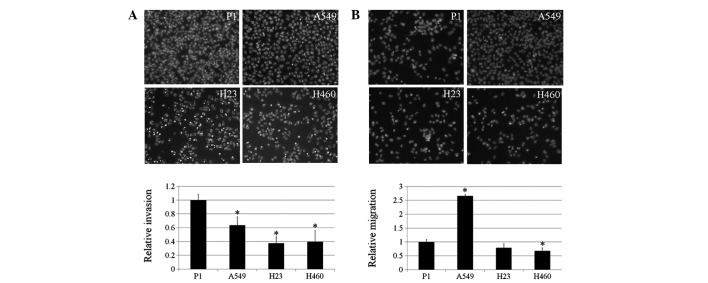
Figure 2.
Invasion and migration of P1, A549, H23 and H460 cells. (A) Lung cancer cells were assessed for their invasive characteristics using Matrigel-coated membranes in Transwell chambers. After 48 h, invaded cells were fixed, stained with Hoechst 33342 and visualized under a fluorescence microscope. Columns represent mean ± SD (n=3), *P<0.05, vs. P1 cells. (B) Cells were assessed for migratory capability. Following 12 h, migrated cells were fixed, stained with Hoechst 33342 and visualized under a fluorescence microscope. Columns represent mean ± SD (n=3), *P<0.05, vs. P1 cells.

