Figure 6.
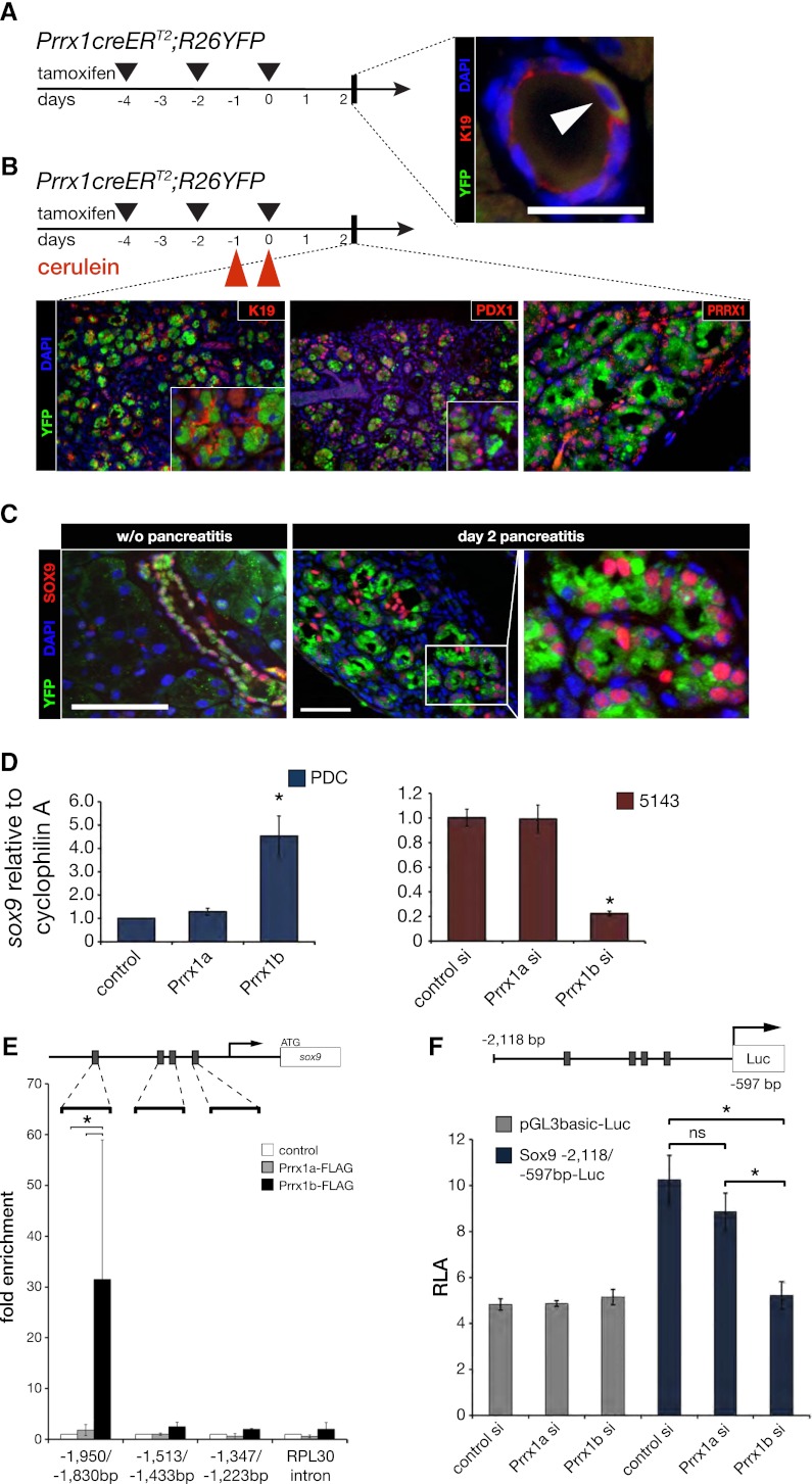
Prrx1b binds the Sox9 promoter. (A) Prrx1creERT2;R26YFP mice. Coimmunofluorescence staining of YFP (green), K19 (red), and DAPI (blue) in representative normal pancreatic intralobular duct (white arrowhead) 48 h after induction with tamoxifen with indicated administration schedule by oral gavage. Bar, 50 μm. (B) Prrx1creERT2;R26YFP mice induced with tamoxifen and treated with cerulein (see the Materials and Methods). Coimmunofluorescence staining of YFP (green); K19, PDX1, or PRRX1 (red); and DAPI (blue) in representative ADM lesions 48 h after cessation of cerulein administration. Each panel shows 200× magnification, and inserts are 400× magnification. (C) Coimmunofluorescence staining in Prrx1creERT2;R26YFP mice of YFP (green), SOX9 (red), and DAPI (blue) in a intralobular duct without cerulein administration or in ADM lesions 48 h after cessation of cerulein administration. Bar, 50 μm. (D) Sox9 expression (qPCR) in PDC-Prrx1a and PDC-Prrx1b cells and Sox9 expression in siRNA to Prrx1a or Prrx1b in 5143 cells (high endogenous Prrx1 level). (E) Schematic diagram of Sox9 upstream 5′ untranslated region. Gray boxes indicate predicted Prrx1-binding sites. Primers were designed for ChIP qPCR flanking the indicated regions. The RPL30 gene intron served as a negative control. There is >30-fold enrichment of Prrx1b binding (Flag immunoprecipitation of PDC-Prrx1b-Flag cells compared with control non-Flag-PDC cells) to the −1950 to −1830 region of the Sox9 gene. There is no enrichment of Prrx1b in regions −1513 to −1433 and −1347 to −1223. Of note, there is no enrichment of Prrx1a to any of the three regions. (F) Prrx1b knockdown (siRNA) reduces Sox9 luciferase reporter activity, whereas Prrx1a knockdown (siRNA) does not. (*) P < 0.05 is statistically significant (Mann-Whitney-Wilcoxon test).

