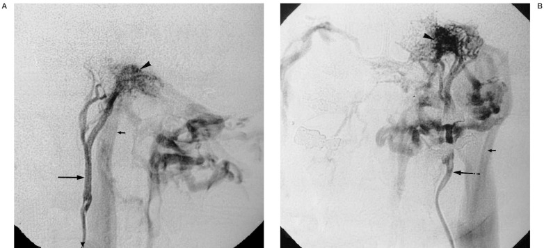
Figure 4.
59-year-old man with suspected right-sided jugular paraganglioma. A) Selective injection of the ascending pharyngeal artery (arrow) in lateral projection shows a highly vascular lesion in the left temporal bone (arrowhead). No real parenchymal stain is seen. Rapid venous drainage in a patent jugular vein is present (small arrow). B) Same in antero-posterior projection, the venous drainage pattern suggests an osteodural arteriovenous malformation.

