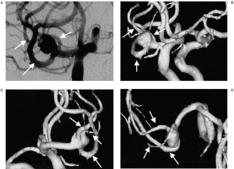
Figure 1.
An aneurysm located at the right middle cerebral artery trifurcation. A) The aneurysm is viewed at right oblique projection on DSA. B-D) On 3D MRA, the aneurysm is viewed at multiple projections on the PC monitor. The relationships between the aneurysm and trifurcating MCA branches (arrows) are more easily demonstrable on the 3D MRA compared with on the DSA image (A) because aneurysm and each arterial branch can be observed at any arbitrary angles.

