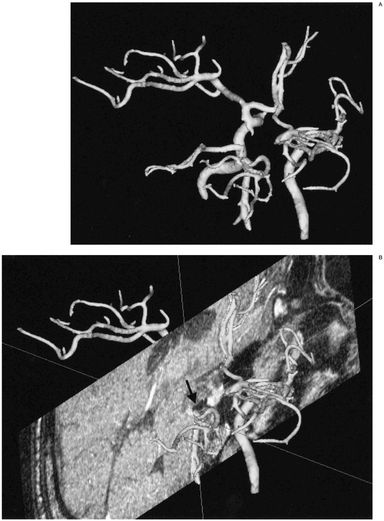
Figure 8.
Background multiplanar MR image assembly. A) A posterior superior view of reconstructed 3D MRA image in a patient with basilar top aneurysm treated by coils one year before. B) 3D MRA image is assembled with the background reconstructed coronal MR image at the level of the basilar top. Previously embolized coil mass is seen as a small area of low signal intensity (arrow) at the top of the basilar artery

