Fig. 4.

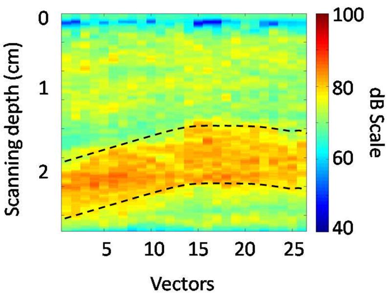
Maximum intensity projection (MIP) subharmonic image obtained from a canine. Note that the abscissa represents the width or the lateral span of the ROI shown in Fig. 3b. The dotted lines delineate the portal vein.

Maximum intensity projection (MIP) subharmonic image obtained from a canine. Note that the abscissa represents the width or the lateral span of the ROI shown in Fig. 3b. The dotted lines delineate the portal vein.