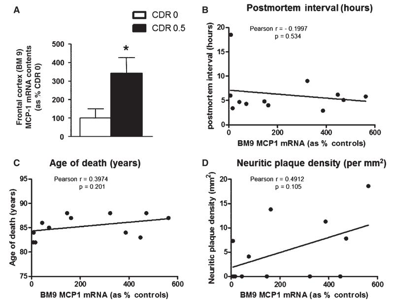
Fig. 5.
Elevated MCP-1 mRNA in frontal cortex (BM9) brain specimens of cases characterized by mild cognitive impairment. (A) Contents of MCP-1 mRNA were assessed by qPCR and normalized to brain contents of human TATA-binding protein mRNA, used as an internal control. Bar graphs represent mean (± SEM) normalized MCP-1 mRNA contents in BM9 of CDR 0 cases (n = 6), expressed as % of MCP-1 mRNA contents in control cases (n = 6). *Student t-test, p < 0.05. B–D) Correlation analysis of BM9 MCP-1 mRNA contents with (B) postmortem interval, (C) age of death, and (D) neuritic plaque density (per mm2). There was no significant correlation between brain MCP-1 mRNA contents and postmortem interval (p = 0.534), age of death (p = 0.201), or neuritic plaque density (p = 0.105).

