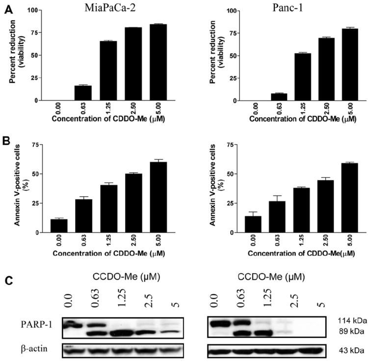
Fig. 1.

CDDO-Me inhibits proliferation and induces apoptosis in pancreatic cancer cells. (A) MiaPaCa-2 and Panc-1 pancreatic cancer cells (1 × 10⁴/well) were treated with CDDO-Me at concentrations ranging from 0 to 5 μM for 72 h in a 96-well microtiter plate in triplicates. Cell viability was measured by MTS assay using CellTiter Aqueous Assay System. (B) Annexin V-FITC binding. Tumor cells were treated with CDDO-Me at 0–5 μM for 48 h and then reacted with 5 μl of annexin V-FITC and 5 μl PI for 30 min and the percentage of annexin V-FITC binding cells was determined by flow cytometry. (C) Cleavage of PARP-1 in cells treated with CDDO-Me was analyzed by immunoblotting. Similar results were obtained in 2 to 3 independent experiments. *P < 0.01 compared to control cells (no CDDO-Me).
