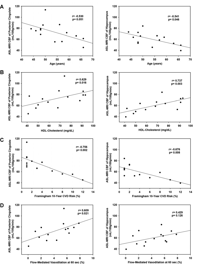
Figure 1.
A. Baseline age correlated inversely with baseline CBF in the posterior cingulate (shown, r=−0.530, p=0.051), hippocampus (shown, r=−0.541, p=0.046), parahippocampus (r=−0.577, p=0.031), thalamus (−0.546, p=0.043), and the cingulate gyrus (r=−0.566, p=0.035). B. Baseline HDL-C correlated directly with baseline CBF in the posterior cingulate (shown, r=0.629, p=0.016), hippocampus (shown, r=0.737, p=0.003), parahippocampus (r=0.722, p=0.004), and thalamus (r=0.607, p=0.021), as well as with global perfusion (r=0.651, p=0.012). C. Higher baseline Framingham 10-year CVD risk correlated with worse baseline CBF in the posterior cingulate (shown, r=−0.756, p=0.002), hippocampus (shown, r=−0.676, p=0.008), parahippocampus (r=−0.712, p=0.004), parietal lobe (r=−0.751, p=0.002), thalamus (r=−0.770, p=0.001), cingulate gyrus (r=−0.712, p=0.004), superior parietal lobe (r=−0.551, p=0.041), and inferior parietal lobe (r=−0.616, p=0.019), as well as with global perfusion (r=−0.719, p=0.004). D. At baseline, FMD at 60 seconds was directly correlated with baseline CBF in the posterior cingulate (shown, r=0.609, p=0.021), thalamus (r=0.701, p=0.005) and cingulate gyrus (r=0.675, p=0.008) and showed a trend toward a positive correlation in the parahippocamus (r=0.512, p=0.061), parietal lobe (r=0.512, p=0.061) and on global perfusion (r=0.516, p=0.059). FMD at baseline did not correlate with baseline CBF in the hippocampus (shown, r=0.429, p=0.126).

