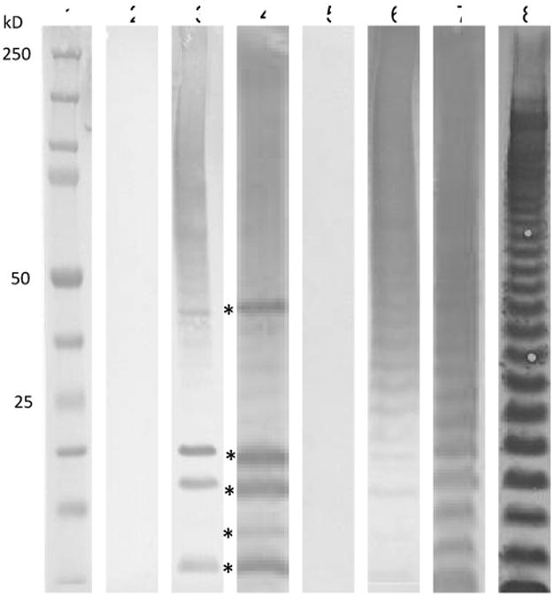
Figure 6.

Anti-MPF antibodies are directed to LPS and membrane proteins. Western blots of MPF (5 μg) with anti-serum from naïve mice receiving 2 MPF injections or from mice infected with Ft Schu S4, 7 days post-infection that received daily gentamicin and 2 MPF injections. Lanes 2-4 show anti-mouse IgG reactivity and lanes 5-7 show anti-mouse IgM reactivity. The 5 most dominant reactive bands in lanes 3 and 4 (*) have apparent masses of 46, 20, 18, 15, and 11 kD respectively. Lane 8 is anti-Ft LVS LPS reactivity. Lanes 2 and 5 contain naïve sera as a control. Lanes 3 and 6 are immune sera from uninfected donor mice. Lanes 4 and 7 are from mice infected with Ft Schu S4 at day 7 post-infection. Lane 1 is a molecular weight marker. Antiserum was pooled from 5 mice for each group.
