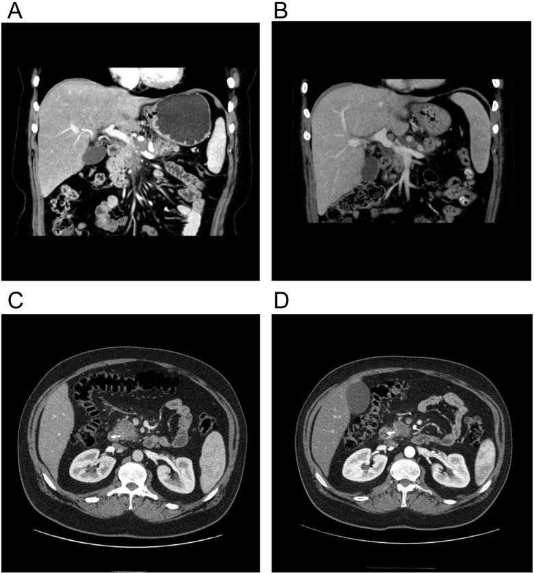
Figure 2.
CT Images in representative responding patients. (A) Pretreatment scan shows extensive mass encircling, and nearly obstructing PV. (B) Posttreatment study shows regression of tumor with widely patent PV. Patient went on to complete resection. (C) Pretreatment CT showing pancreatic head mass. (D) Posttreatment CT shows response of tumor in pancreatic head and retraction from superior mesenteric vein. Patient went on to complete resection. CT, computed tomography; PV, portal vein.

