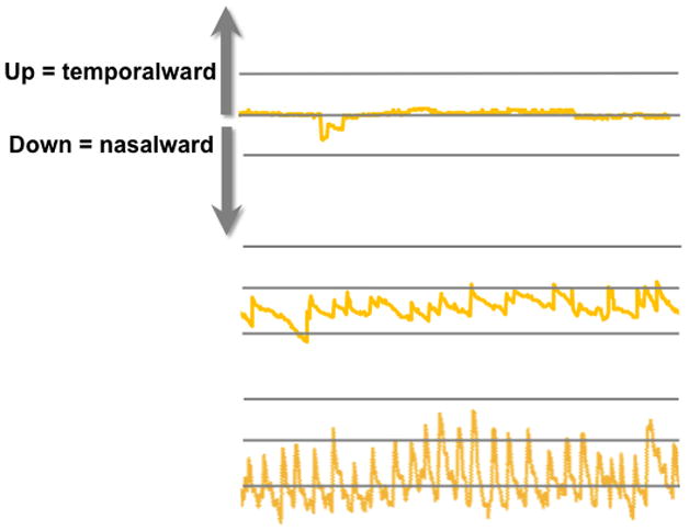
Figure 12.

Eye position records derived from the Nidek MP-1 for the 3 anisometropic children shown in Figure 10.(Birch et al., 2012) The top trace shows eye position for the child with normal fixation stability (Figure 10, left); overall, fixation is accurate, with only microsaccades and a brief saccadic oscillation. The middle trace shows the typical anisometropic child (Figure 10, middle); it shows the classic waveform of FMNS with slow drifts nasalward, and rapid re-fixating temporalward saccades. The bottom trace shows the anisometropic child with extreme fixation instability (Figure 10, right); high frequency, large amplitude FMNS is evident.
