
Figure 9.
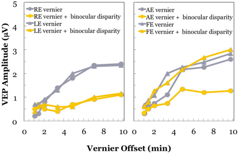
VEP responses to monocular oscillating vernier offsets recorded from a child with normal vision (left) and an amblyopic child (right) with 0 or 5 min standing binocular disparity.(Fu et al., 2006) For the child with normal vision, vernier responses are similar for each eye (grey symbols). The addition of a standing binocular disparity results in decreased VEP amplitude, consistent with fusional suppression. The amount of suppression is similar regardless of which eye views the vernier target and which views the static, disparate stimulus (yellow symbols). For the amblyopic child (right), the amblyopic eye has slightly, but consistently lower, amplitude vernier responses than the fellow eye. When the standing disparity is introduced to the fellow eye, fusion suppression is observed (yellow circles). However, when the standing disparity is introduced in the amblyopic eye, no fusional suppression is observed (yellow triangles). Fusion suppression is asymmetric in the amblyopic child.