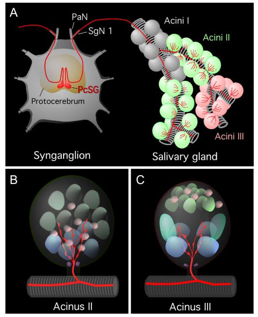
Figure 7.
A: Simplified schematic diagram showing the axonal projection from the PcSG neuron to the salivary glands. Different colors of nuclei in B and C indicate different types of cells based on their size and shape in the confocal image (purple, neck cells; blue, basal granular cells; green, apical granular cells; pink, epithelial cells in B; purple, neck cells; blue, basal granular cells; aqua blue, basal cells; greenish gray, apical cells; pink, epithelial cells in C. Note that only acini II and III are innervated, whereas MIP/SIFamide innervation was never observed in acini I. Four or five axonal branches in acini II and three or four branches in acini III are in contact with the basal cells that have large, round nuclei. See nomenclature of peptidergic neurons and list of Abbreviations.

